Epsom and Ewell Times

Current
ISSN 2753-2771

The Hidden Link Between Metabolic Syndrome and Joint Pain

Many people have heard of metabolic syndrome as a risk factor for heart disease, stroke, and diabetes — but it’s now becoming clear that it also takes a serious toll on our bones, joints, and muscles.

Metabolic syndrome is a cluster of conditions, including obesity, high blood pressure, raised blood sugar, high triglycerides, and low levels of “good” cholesterol (HDL). Affecting nearly one in three adults in the UK, it’s driven largely by sedentary lifestyles and poor diet.

But beyond its impact on the heart, metabolic syndrome causes long-term inflammation in the body, which in turn affects musculoskeletal health in several key ways:

  • Joint Pain & Arthritis: Chronic inflammation from visceral fat can damage cartilage and accelerate the development of osteoarthritis, particularly in the knees and hips.
  • Tendon Problems: Conditions like Achilles tendinopathy and shoulder pain are more common in people with metabolic syndrome. High blood sugar can stiffen tendons, making them prone to injury.
  • Bone Health: There’s a strong link between metabolic syndrome and reduced bone density. Inflammation and insulin resistance disrupt normal bone repair, increasing the risk of fractures and osteoporosis.

The condition also interferes with the body’s ability to heal and maintain tissues, meaning injuries can linger and become chronic.

The good news? Physiotherapy and regular exercise play a crucial role in managing the effects of metabolic syndrome on the musculoskeletal system. By improving mobility, reducing inflammation, and supporting healthy weight loss, targeted movement and rehab strategies can make a real difference.

So if you’re living with joint or tendon pain and also have risk factors for metabolic syndrome, it might be time to take a more holistic view — and seek advice from a physiotherapist or your GP.

The Role of Physiotherapy and Exercise

Despite its challenges, metabolic syndrome’s effects on musculoskeletal health can be mitigated through physiotherapy and exercise.

1. Exercise as an Anti-Inflammatory Intervention

Regular exercise reduces chronic inflammation by promoting anti-inflammatory cytokines such as IL-10 while lowering pro-inflammatory markers. A study in Diabetes Care (2014) showed that aerobic exercise significantly reduced CRP and TNF-α levels in individuals with metabolic syndrome.

Weight loss through exercise reduces visceral fat, a major source of pro-inflammatory cytokines, easing joint pain and improving musculoskeletal function.

2. Physiotherapy for Joint Pain and Tendinopathies

Physiotherapy plays a key role in managing musculoskeletal conditions related to metabolic syndrome. Personalized exercise programs focusing on strength, flexibility, and joint stability help manage OA and prevent further joint damage.

For tendinopathies, physiotherapists recommend strengthening exercises, which promote tendon healing and reduce pain. A British Journal of Sports Medicine (2017) study found that eccentric exercises significantly improved function and reduced pain in Achilles tendinitis, even in individuals with metabolic syndrome.

Additionally, physiotherapists provide guidance on body mechanics and joint protection strategies, reducing strain on joints and tendons during daily activities.

3. Bone Health and Resistance Training

Resistance training is essential for bone health in individuals with metabolic syndrome. Weight-bearing exercises, such as strength training and resistance bands, stimulate bone formation and help maintain density. A Journal of Bone and Mineral Research (2018) study found that resistance training improved BMD in postmenopausal women with metabolic syndrome, reducing osteoporosis and fracture risk.

Balance and coordination exercises can also be incorporated to prevent falls, particularly for individuals with weakened bones.

Conclusion: Addressing Metabolic Syndrome for Better Musculoskeletal Health

Metabolic syndrome significantly increases the risk of osteoarthritis, tendinopathies, and osteoporosis due to chronic inflammation and tissue dysregulation. However, these negative effects can be mitigated through physiotherapy and regular exercise.

By reducing inflammation, improving metabolic health, and promoting tissue repair, exercise and physiotherapy enhance musculoskeletal function and overall well-being. Individuals with metabolic syndrome can benefit from tailored exercise programs and physiotherapy interventions to manage joint pain, prevent injuries, and maintain strong bones and healthy tissues.

Luke Schembri, Advanced Physiotherapy Practitioner

http://www.genuinephysio.com/

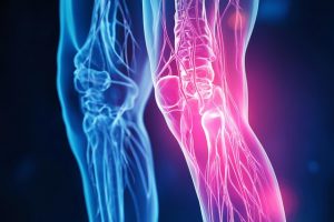
Image by Tung Lam from Pixabay


Hip pain explained

Hip pain is a common complaint, particularly among middle-aged and older adults. Two frequent causes are Hip Osteoarthritis (OA) and Greater Trochanteric Pain Syndrome (GTPS). While these conditions share some symptoms, they differ in causes, treatment, and long-term outcomes. Understanding these distinctions is key for accurate diagnosis and management.

What is Hip Osteoarthritis?

Hip osteoarthritis is a degenerative joint disease characterized by age-related changes in the cartilage of the hip joint. The hip’s ball-and-socket structure relies on cartilage for smooth movement, but this can wear down over time, leading to pain, stiffness, and decreased function.

Signs and Symptoms of Hip OA:

  • Pain in the Groin or Thigh: Pain is typically deep in the groin, buttock, or thigh, worsening with activity and improving with rest.
  • Stiffness: Morning stiffness lasting less than 30 minutes is common, especially after inactivity.
  • Reduced Range of Motion: Difficulty with daily activities such as putting on socks or shoes.
  • Crepitus: A grinding sensation during movement due to rough joint surfaces.
  • Functional Limitations: Challenges with walking, climbing stairs, or standing up from a seated position.

What is Greater Trochanteric Pain Syndrome?

Greater Trochanteric Pain Syndrome (GTPS) involves pain over the lateral hip, specifically around the greater trochanter, the bony prominence on the hip’s outer side. Unlike OA, GTPS stems from issues in the soft tissues, such as the gluteal tendons and bursae.

Signs and Symptoms of GTPS:

  • Lateral Hip Pain: Pain over the outer hip, often radiating down the thigh and exacerbated by lying on the affected side or climbing stairs.
  • Tenderness: Pain upon palpation of the greater trochanter.
  • Pain with Activity: Aggravated by repetitive movements or prolonged standing.
  • Night Pain: Discomfort when lying on the affected side, disrupting sleep.
  • Muscle Weakness: Weakness in the hip abductors, affecting gait and mobility.

Key Differences Between Hip OA and GTPS

  1. Location of Pain

    • Hip OA: Pain is typically deep in the groin, buttock, or thigh, potentially radiating to the knee.
    • GTPS: Pain is localized to the lateral hip, radiating down the outer thigh but rarely affecting the groin.

  2. Underlying Pathology

    • Hip OA: A degenerative joint disease involving cartilage degradation.
    • GTPS: A soft tissue condition involving inflammation or degeneration of the gluteal tendons and bursae.

  3. Risk Factors

    • Hip OA: Aging, obesity, joint injuries, genetics, and repetitive hip loading.
    • GTPS: More common in peri- and post-menopausal women due to hormonal changes affecting tendon health, as well as altered biomechanics like gait changes or muscle weakness.

  4. Impact of Hormones on Soft Tissue Health Hormonal changes during menopause reduce tendon elasticity, increasing the risk of GTPS. In contrast, OA primarily results from mechanical factors and cartilage degradation.

Management and Treatment

Hip Osteoarthritis:

  • Exercise and Physiotherapy: Low-impact activities like swimming or cycling help maintain joint mobility and muscle strength.
  • Weight Management: Reducing body weight can alleviate stress on the hip joint.
  • Medication: NSAIDs are commonly used for pain and inflammation.
  • Joint Injections: Corticosteroid injections under ultrasound guidance can provide temporary relief and aid diagnosis in complex cases.
  • Surgery: Total hip replacement may be necessary in severe cases.

Greater Trochanteric Pain Syndrome:

  • Activity Modification: Reducing activities that exacerbate symptoms, such as prolonged standing or lying on the affected side.
  • Physiotherapy: Strengthening the hip abductor muscles and improving flexibility is key.
  • Shockwave Therapy: Effective in some cases for promoting tissue healing and reducing pain.
  • Corticosteroid Injections: These can temporarily reduce inflammation for patients unresponsive to conservative treatment.
  • Surgery: Rarely required but an option for severe or unresponsive cases.

Conclusion

Hip Osteoarthritis and Greater Trochanteric Pain Syndrome are distinct conditions causing hip pain, with different causes, symptoms, and treatments. While OA is a degenerative joint condition affecting cartilage, GTPS is a soft tissue disorder involving the tendons and bursae around the greater trochanter. Accurate diagnosis is essential for effective management, and consulting a healthcare professional is critical for those experiencing hip pain.

For more information see www.genuinephysio.com

Image: Man with hip pain. Credit Towfiqu Barbhuiya


Frozen Shoulder Link to Menopause

Frozen shoulder, also known as adhesive capsulitis, is a painful condition that restricts movement in the shoulder joint due to adhesion and inflammation in the joint capsule. While this condition can affect anyone, it is particularly common in women aged 40 to 60, with many cases coinciding with menopause.

What Is Frozen Shoulder?

Frozen shoulder is characterised by pain and stiffness in the shoulder joint. It progresses through two main stages: the painful stage, where movement becomes uncomfortable and often disrupts sleep, and the stiffness stage, where shoulder mobility is severely restricted. The exact cause isn’t always clear, but there are certain risk factors.

The Menopause Connection

Frozen shoulder tends to occur more frequently in women, especially during menopause. Hormonal changes, particularly declining oestrogen levels, can impact connective tissues, making them more prone to inflammation and stiffness. This hormonal shift may partly explain why women in midlife are at higher risk of developing the condition.

Signs and Symptoms

Key symptoms of frozen shoulder include:

  • Pain: A deep, aching pain that worsens with movement. Night pain is common.
  • Stiffness: As the condition progresses, shoulder movements become more restricted, making daily activities like dressing or reaching overhead challenging.
  • Limited Range of Motion: Both active and passive movements are affected.

Risk Factors for Frozen Shoulder

  • Age and Gender: Women between 40 and 60, especially those going through menopause, are at higher risk.
  • Diabetes: Those with diabetes are more susceptible and often experience a more prolonged course of frozen shoulder.
  • Thyroid Disorders: Hypothyroidism and other thyroid issues are linked to a higher likelihood of developing frozen shoulder.
  • Immobilisation: Lack of movement after an injury or surgery can trigger the condition.

Treatment and Management Options

Physiotherapy: The Gold Standard

Physiotherapy is the cornerstone and is supported by the National Institute for Health and Care Excellence (NICE). It includes a combination of manual therapy, exercise, and education to relieve symptoms and improve shoulder function.

  • Manual Therapy: Techniques like joint mobilisation and stretching are used to reduce stiffness and increase mobility.
  • Exercise Therapy: A personalised exercise plan can help restore shoulder movement and build strength. Early in the painful stage, exercises are gentle, with intensity increasing as the shoulder improves.
  • Patient Education: Understanding frozen shoulder, its natural progression, and expected recovery time is vital. Many patients worry about the severity of their pain, especially during menopause, but education can reassure them that frozen shoulder is manageable and temporary.

Injection Therapy: Corticosteroids and Hydrodilatation

For those whose pain severely limits rehabilitation, injection therapy can be an effective complement to physiotherapy.

  • Corticosteroid Injections: These are most useful in the early painful stage, providing significant pain relief and reducing inflammation. This helps patients participate more actively in physiotherapy.
  • Hydrodilatation: This procedure involves injecting a saline solution, often combined with a corticosteroid, into the joint capsule to stretch it and improve mobility. It’s typically used during the stiffness phase.

Can Frozen Shoulder Be Prevented?

While not always preventable, staying active and maintaining shoulder mobility can reduce the risk of developing frozen shoulder, particularly during menopause when hormonal changes increase vulnerability. If you experience early signs of stiffness or discomfort, seeking physiotherapy promptly can prevent the condition from worsening.

Role of Menopause in Frozen Shoulder Recovery

Due to hormonal changes, women going through menopause may experience a longer recovery time. However, with early intervention, including physiotherapy and, if necessary, injection therapy, most women see significant improvements within one to three years. Managing underlying conditions like diabetes or thyroid issues can also speed up recovery.

Conclusion

Frozen shoulder is a painful and limiting condition, but with the right treatment, particularly physiotherapy, most people can regain their shoulder mobility over time. For women experiencing menopause, the added risk makes it important to be proactive in addressing early symptoms. Whether through manual therapy, tailored exercises, or injection therapy, effective treatment can help you regain shoulder function and return to your regular activities.


Local women’s health specialist on GTPS

Greater Trochanteric Pain Syndrome (GTPS) is a prevalent condition, particularly affecting peri- and post-menopausal women. It causes significant discomfort in the soft tissue on the outside of the hip and can severely limit mobility. This article discusses the signs, causes, and management of GTPS, with a focus on why it predominantly affects women undergoing hormonal changes.


Luke Schembri is an Advanced Physiotherapy Practitioner working within the NHS, while also offering care to a limited number of private patients from his home. Born and raised in Epsom, he has always lived near Epsom Downs, which inspired his initial interest in the horseracing industry where he began his physiotherapy career. Luke is dedicated to delivering evidence-based, high-quality care to his local community. In addition, he authors a fortnightly blog that addresses health and wellbeing topics, particularly aimed at individuals over the age of 40.


What is Greater Trochanteric Pain Syndrome?

GTPS is characterized by pain and tenderness over the greater trochanter, a bony area on the outside of the hip. The condition involves inflammation of the gluteal tendons or bursa, and although it was once called trochanteric bursitis, the term GTPS is now preferred as it covers a broader range of tendon-related disorders.

Signs and Symptoms

The primary symptom of GTPS is pain over the lateral hip, which can radiate down the outer thigh. Key symptoms include:

– Lateral Hip Pain: Often worsened by activities like lying on the affected side, walking, or sitting in low chairs.

– Tenderness Over the Greater Trochanter: Touching this area typically causes discomfort.

– Pain with Activity: Repetitive movements, such as climbing stairs, exacerbate the pain.

– Night Pain: Pain at night is common, particularly when lying on the affected side.

– Weakness or Stiffness: Some may experience hip weakness or stiffness, affecting their gait.

Why is GTPS Most Common in Peri- and Post-Menopausal Women?

GTPS disproportionately affects women in peri- and post-menopause due to hormonal changes and mechanical stress on the hip. Here are the primary reasons:

Hormonal Changes

The decline in oestrogen during menopause impacts the health of tendons, ligaments, and muscles. Oestrogen plays a critical role in maintaining soft tissue integrity, and reduced levels lead to:

– Decreased Collagen Production: Collagen is essential for tendon strength and elasticity, and its decline makes tendons more prone to injury.

– Increased Tendon Stiffness: Lower oestrogen increases tendon stiffness, contributing to gluteal tendinopathies.

– Altered Pain Perception: Hormonal changes during menopause can heighten sensitivity to pain, worsening GTPS symptoms.

Mechanical Load and Changes

Other factors contributing to GTPS in menopausal women include:

– Increased Load on the Hip: Weight gain during menopause places more stress on the hip joints and surrounding tissues.

– Reduced Physical Activity: Lower activity levels can cause muscle weakness, altering hip mechanics and increasing the likelihood of tendon injuries.

– Altered Gait: Pain-induced changes in walking patterns can further strain the hip and lead to GTPS.

Treatment and Management of GTPS

Treatment for GTPS involves conservative methods, physiotherapy, and sometimes more invasive procedures, following the National Institute for Health and Care Excellence (NICE) guidelines, which recommend starting with non-invasive options.

1. Conservative Management

   – Rest and Activity Modification: Reducing activities that worsen symptoms, such as standing for long periods or lying on the affected side, is crucial. Using a pillow between the knees when sleeping may help alleviate pressure on the hip.

   – Non-Steroidal Anti-Inflammatory Drugs (NSAIDs): These medications can reduce pain and inflammation and are often recommended as initial treatment.

2. Physiotherapy

   Physiotherapy plays a key role in treating GTPS and involves:

   – Strengthening Exercises: Targeted exercises to strengthen the hip abductors, particularly the gluteus medius and minimus, improve hip stability and reduce strain on the tendons.

   – Manual Therapy: Techniques such as deep tissue massage and myofascial release can help relieve muscle tightness and provide short-term pain relief.

   – Education and Advice: Physiotherapists offer valuable guidance on managing activities and loading the hip joint to prevent further injury.

3. Shockwave Therapy

   Extracorporeal shockwave therapy (ESWT) is a non-invasive option that promotes healing by delivering shockwaves to the affected area. Some studies show positive outcomes for GTPS patients treated with ESWT.

4. Corticosteroid Injections

   For patients who do not respond to conservative treatments, corticosteroid injections may be used to reduce inflammation. However, repeated injections can weaken the tendons, so they are not a long-term solution.

5. Surgery

   Surgery is rare and reserved for severe cases where other treatments have failed. Options include removing the inflamed bursa or repairing the tendons.

Conclusion

Greater Trochanteric Pain Syndrome is a common condition that primarily affects peri- and post-menopausal women. Hormonal changes and mechanical stress on the hip contribute to its development. Effective treatment usually involves conservative management, physiotherapy, and sometimes shockwave therapy or injections. Early diagnosis and a tailored treatment plan can help alleviate symptoms, improve function, and enhance the quality of life for those affected by GTPS.

Page 1
© 2021-2025. No content may be copied without the permission of Epsom and Ewell Times Ltd.
Registered office: Upper Chambers, 7 Waterloo Road, Epsom KT19 8AY